
Dr. Mark Bekhit, Radiologist · Last updated August 2026
Short answer: Pelvic MRI is not the primary test for diagnosing endometriosis. Its clearest role is in secondary care, where it maps suspected deep endometriosis and whether nearby pelvic organs could be involved. It is a mapping tool, not a simple yes-or-no test, and it cannot rule endometriosis out on its own. Follow your appointment instructions, then ask your referrer: “What are you hoping this MRI will clarify?”
What can pelvic MRI show when endometriosis is suspected?
Pelvic MRI is most useful when the clinical team needs a map of suspected deep endometriosis. In secondary care, it can “catalogue” where suspected deep disease is and whether it involves another pelvic organ. It is not the first or only diagnostic test.
The 2020 New Zealand consensus defines “deep” as disease extending more than 5 mm beneath the lining of the pelvis, or involving or distorting the bowel, bladder, ureter or vagina. Those named structures explain why a pelvic MRI report can read like an anatomical map rather than a simple positive-or-negative result.
This mapping can be useful before a specialist discussion because surgery for suspected deep disease can require particular expertise and, at times, input from more than one specialty. It is still the specialist’s job to put the images beside your symptoms, examination, previous imaging, response to treatment and priorities. MRI supplies part of that picture; it does not choose the treatment.
A useful question for your referrer: “Which part of my symptoms or previous imaging do you want this MRI to clarify?” The answer tells you whether the purpose is to map suspected deep disease, look at possible organ involvement or assess another pelvic finding.
If you are still unsure why MRI rather than another test was requested, the MRI-versus-CT booking guide explains the broader differences between the two scans. Your own referral remains the source of truth for this appointment.
Can a normal pelvic MRI rule out endometriosis?
No. A normal pelvic MRI is not a definitive all-clear for endometriosis. MRI should not be used as the primary diagnostic tool, and definite diagnosis requires laparoscopy. Assessment and management draw on the clinical history, examination and other investigations, not one MRI result alone.
Symptoms still deserve attention even when a report does not identify deep disease. Possible features of endometriosis include painful periods, pelvic pain, deep pain during or after sex, bowel symptoms, urinary symptoms and difficulty becoming pregnant. These symptoms are not specific to one condition, so they guide assessment rather than prove a diagnosis.
The amount of pain does not reliably tell you how extensive the disease is. A report that describes little or no deep disease therefore should not be used to dismiss severe symptoms, and a more extensive imaging finding does not by itself reveal how much pain a person has.
If symptoms remain uncontrolled after primary-care management, referral to secondary-care gynaecology is appropriate. That is a pathway decision for your healthcare team, not a conclusion that follows automatically from the scan. The useful follow-up is to ask what the MRI did answer, what it did not explain and what part of the assessment comes next.
Why might the report mention other organs or adenomyosis?
The radiologist’s report describes what is visible on the images and where it is located. For suspected deep endometriosis, MRI includes looking for possible involvement of other pelvic organs. The report can also describe a different or additional finding, such as adenomyosis. An MRI appearance suggesting adenomyosis does not exclude endometriosis occurring at the same time.
One line of a report does not determine a treatment plan. Ask the person discussing it with you to show which finding answers the referral question and which part remains uncertain.
How should I prepare for a pelvic MRI?
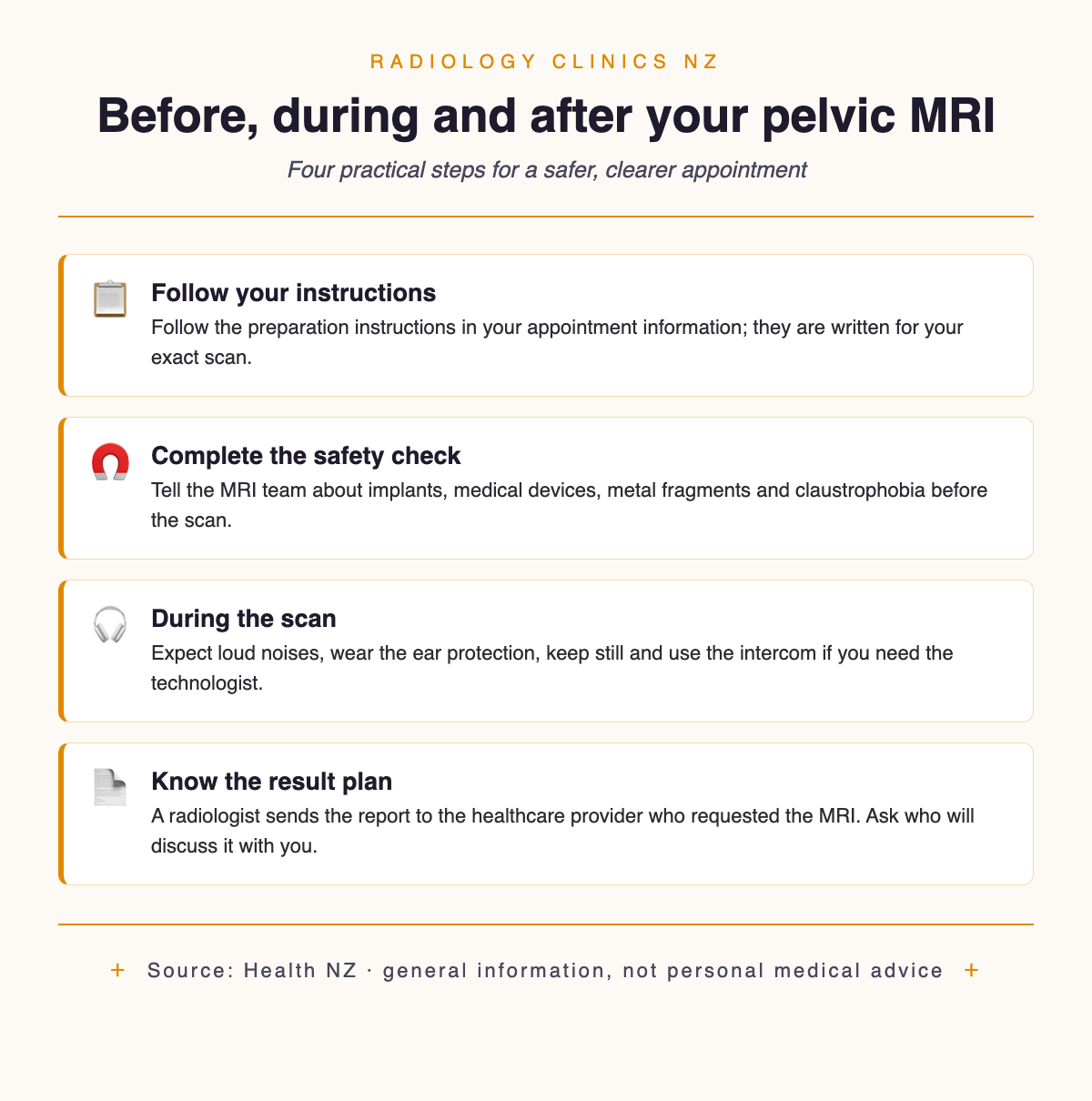
Start with the preparation instructions sent for your MRI appointment. They are more reliable than a generic checklist because the imaging service knows the exact examination requested. The imaging appointment checklist can help with documents and practical items, but it does not replace your booking instructions.
MRI uses a powerful magnet, so the safety questionnaire matters. Tell the team about implanted medical devices, metal in your body and any history of metal fragments before you enter the scanner room.
If enclosed spaces are difficult for you, contact the service before scan day. Coping techniques or a sedative can help some people with claustrophobia. If sedation is arranged, for 24 hours you should not drive, operate machinery, drink alcohol, sign legal documents or travel alone on public transport. Arrange your transport before the appointment rather than discovering that restriction afterwards.
Some MRI examinations use an injection of gadolinium contrast; others do not. Your appointment information or MRI team will tell you whether it is planned for this scan and complete the relevant safety checks. For a fuller explanation of contrast questions, use the contrast allergies guide and ask specifically, “Is contrast planned for my pelvic MRI, and is there anything in my history you need to check?”
Before you agree to the scan
✓ Ask for enough information to understand the purpose, main benefits, risks and alternatives.
✓ Ask for an honest answer to anything that is unclear.
✓ Remember that you can refuse or withdraw consent.
✓ Ask about a support person; this right is qualified where safety or another person’s rights would be compromised.
These are New Zealand rights, not favours. Rights 6, 7 and 8 of the Code of Health and Disability Services Consumers’ Rights cover information, informed choice, refusal or withdrawal of consent, and support people. If you need an interpreter, NZSL or information in another format, the radiology communication support guide has an exact booking script.
What happens during and after the scan?
MRI uses a strong magnet and radio waves to create detailed images. It does not use ionising radiation—the type used in X-rays and CT. MRI examinations commonly take 15–90 minutes, depending on the body part (timing guidance checked August 2026). Your appointment information is the better guide for how long to allow for this pelvic scan.
The scanner is noisy, so the team gives you earplugs or headphones. You need to keep still while images are being collected, and you can speak with the MRI technologist through the intercom.
Tell the technologist if pain makes one position difficult or if you need to pause. The team can explain what is possible without promising that every scan can be changed in the same way. Clear communication matters more than trying to tolerate a problem silently.
After the images are complete, a radiologist reviews them and writes a report. The report goes to the healthcare provider who requested the examination, and that provider communicates the result. Ask when and how the result will be discussed with you.
Before leaving, ask who owns the next step and when you should expect contact. The first radiology scan questions provide a broader list if this is your first imaging appointment.
What should I ask when the MRI report is discussed?
Use the report to organise the conversation, not replace it. Management choices take account of symptoms, clinical history, disease extent, other health conditions, cultural considerations and your own priorities, including pain control and fertility. Treatment is also shaped by fertility plans and personal preference.
Six questions for the follow-up
✓ What question was the MRI intended to answer?
✓ Did it describe suspected deep endometriosis or involvement of another organ?
✓ Did it show adenomyosis or another possible explanation?
✓ What does the scan not explain about my symptoms?
✓ How does this result change my options, given my pain and fertility priorities?
✓ Do I need gynaecology review or an opinion from a team with deep-endometriosis expertise?
If suspected deep endometriosis is being considered for surgery, care should involve a gynaecologist with special surgical expertise supported by a multidisciplinary team. That does not mean every person with an imaging finding needs surgery. It tells you what expertise to ask about if a surgical option is being discussed.
You also have the right to honest and accurate answers and enough information for an informed choice. A patient-centred radiology appointment should leave you clear about who will explain the result and what happens next. If you need to locate a service, use the guide to finding a radiology clinic or search the New Zealand clinic directory; MRI still requires a referral.
Frequently asked questions
How long does a pelvic MRI for endometriosis take?
MRI examinations commonly take 15–90 minutes, depending on the body part being examined (timing guidance checked August 2026). Your appointment information is more specific because it reflects the images planned for your pelvic scan. Allow extra time for check-in and safety screening if the service asks you to arrive early.
Does pelvic MRI use radiation?
No. MRI uses a strong magnet and radio waves, not ionising radiation. The magnetic field is the reason staff screen carefully for metal, implants and medical devices before you enter the scanner room.
Will I need contrast?
Some MRI examinations use gadolinium contrast, but that does not mean every pelvic MRI does. Follow the instructions for your booked examination and ask whether an injection is planned. The MRI team completes the relevant safety check when contrast is part of the protocol.
Can I self-refer for pelvic MRI in New Zealand?
No. MRI requires a referral and can be provided through public or private services. The requester supplies the clinical question that helps the radiology team plan and interpret the examination.
Can I bring a support person?
You have the right to one or more support people unless their presence could compromise safety or unreasonably infringe another person’s rights. Ask before the appointment how that works around the MRI safety zone and during image collection.
Sources and review
This page was clinically reviewed against New Zealand sources on 6 August 2026. The disease-specific MRI role comes from the Ministry of Health’s 2020 Diagnosis and Management of Endometriosis in New Zealand, which is a consensus view rather than a formal clinical guideline. General scan information comes from Health NZ’s MRI page and its scans and X-rays overview. Symptom and management context was checked against Health NZ and Healthify. Consent and support-person statements come from the Health and Disability Commissioner’s Code. Your appointment provider supplies the exact preparation and contrast instructions for your booked examination.
This article is general information, not personal medical advice. Ask your referrer which service is available, and ask the clinic whether public funding or ACC cover applies and what you will need to pay.

Author: Dr. Mark Bekhit
Dr Mark Bekhit (MBChB, FRANZCR) is a musculoskeletal radiologist practising in Brisbane and an AHPRA-registered specialist in diagnostic radiology (MED0002920947). He spends as much time building software as reading scans: he founded Imaging Finder (Australia) and Radiology Clinics NZ to make imaging easier to find, and builds radiology dictation and pain-tracking tools with one aim — better patient outcomes and a smoother reporting workflow. Outside the reading room he's usually training, bringing the same performance mindset to imaging.
Sources, method, and medical context
This article is published by Radiology Clinics NZ and reviewed for medical-adjacent accuracy by Dr. Mark Bekhit, radiologist. It is general information only and does not replace advice from your own doctor, referrer, clinic, insurer, or Health New Zealand service.
Official references
- Health New Zealand: scans and X-rays
- Health New Zealand: community-referred radiology service
- Radiology Clinics NZ: how the directory is built
Related directory pages
Radiology clinics directoryMRI clinicsBrowse by cityHow this directory is built
Last editorial review: 2026-06-30.
Leave a Reply