The evening you find a lump is a long one, and it usually ends with a decision about who to ring in the morning.
The short answer A lump or change is a symptom, and symptoms do not go through the free screening programme — having no symptoms of breast cancer is one of its own conditions. See your own healthcare provider as soon as possible instead. They will examine you and may refer you for a mammogram or an ultrasound; ultrasound is more likely to be used if you are young or have dense breast tissue. Most breast lumps are not cancer. Do not wait for your next screening mammogram.
Start with your own doctor, not the screening programme
Many people spend that first evening deciding to ring the free breast screening number. This is the part that trips people up, so it is worth being blunt. BreastScreen Aotearoa is a screening programme; it looks for cancer in people who feel completely well. One of the stated conditions for a free two-yearly mammogram is that you have no symptoms of breast cancer. Once you have noticed something, you are no longer in that group. Your GP, nurse practitioner or usual healthcare provider is the right door.
Go early rather than politely. The reason is not that a lump is usually sinister; it usually is not. The reason is that the only way to find out is to look. Half of all breast cancers are found because someone noticed a change and showed their healthcare provider (Healthify, accessed September 2026). That number exists precisely because people acted on something small.
Changes worth an appointment. A new lump in the breast or armpit; a change in breast shape or size; skin dimpling, puckering or thickening; a nipple that turns inwards, ulcerates or newly itches; nipple discharge or bleeding; persistent redness; or breast pain that does not go away. A rash or reddening on one breast only belongs on the list too.
Any of these means an appointment rather than a wait-and-see — including when your next screening mammogram is already booked.
A clear screening mammogram six months ago does not change that either; cancers can appear between rounds, and there is a name for it — an interval cancer. None of which means expecting bad news. The common causes of a breast lump are benign: normal cyclical changes, a fibroadenoma, a cyst, fat necrosis, a lipoma. You just cannot tell them apart by feel, at home, at ten at night. Neither can a radiologist, not without the images.
Which scan you are likely to have
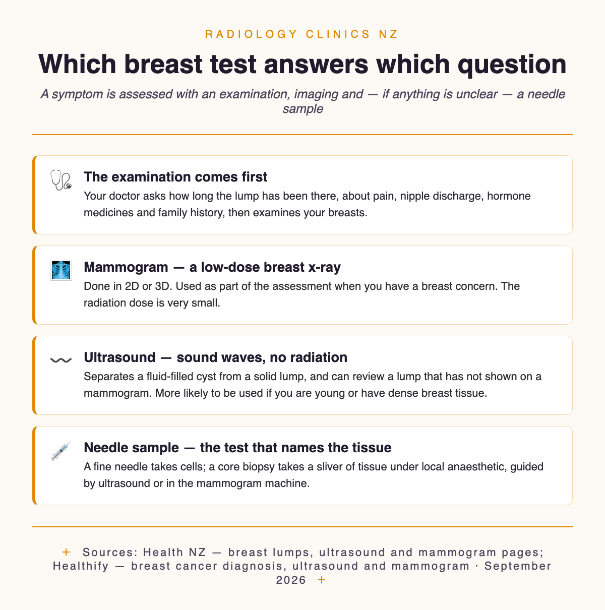
There is no single “breast scan”. A symptom is worked up with a combination of methods — a physical examination, imaging such as a mammogram and ultrasound, and, if needed, a biopsy and testing of the cells. Your doctor picks the starting point from your age, your breast tissue and what they can feel. It is a clinical decision, not a menu.
Two rules are worth knowing. A mammogram may be asked for as part of the assessment when you have a breast concern, and ultrasound is sometimes used as well as, or instead of, a mammogram to check for breast lumps — more likely if you are young or have dense breast tissue. The two are not rivals; they answer different questions, which is why they are so often done together.
The most useful thing I can tell you as a radiologist is what a normal mammogram does not settle. Occasionally a lump that can be felt is not seen on a mammogram, and such a lump should not be ignored; other tests need to be done. In women aged 45 to 69, about one or two out of every ten cancers are not found by mammograms (Health NZ, accessed September 2026). So if you can still feel something and the x-ray was clear, the sentence you want from your doctor is not “the mammogram was fine”. It is “the mammogram was fine, and here is what we are doing about the lump.”
Where to go, and what to ask when you book
Once the referral is written you often have some say in where it goes, and breast imaging is not offered at every radiology branch. The mix differs even between sites in the same network. Start with our mammography clinics hub, which groups the New Zealand clinics filed under mammography in our directory. Those listings currently sit in North Island regions, so if you are further south go instead to the directory search for breast ultrasound, which also reaches Wellington, Taranaki, Canterbury, Otago and Southland. That search matches either word, so it mixes dedicated breast clinics with general ultrasound practices. Each listing opens with the address, phone number and website.
The short service line under a listing name is a summary, and it can lag what a clinic actually offers. The four examples below were checked against each provider’s own current page on 6 September 2026, and are not a ranking, a recommendation or a complete list.
Worth sorting before the appointment
✓ Wear a separate top and bottom: you undress from the waist up for a mammogram and for a breast ultrasound.
✓ Skip the deodorant. Creams, lotions, deodorant, perfume, body oils, powders and sunscreen on the breasts or armpits can affect the x-ray image.
✓ Bring any earlier reports you have at home — comparing with previous results helps the radiologist.
✓ Ask whether the branch has 3D mammography, since not all providers offer it and there may be a cost.
Chase the appointment if it does not appear: your provider requests the test and you should get a letter or a call with the time and place, so contact your provider or hospital doctor if neither arrives. And if the symptom changes while you wait — a lump getting bigger, say — go back to your main healthcare provider so the priority can be reassessed. A waiting list is not fixed in stone.
What actually happens at the appointment
It starts with talking, not scanning. Your provider will ask how long the lump has been there, about breast pain and nipple discharge, when your last period was, whether you take hormone medication such as the contraceptive pill or menopause hormone therapy, whether you have had lumps before and whether breast disease runs in your family — then examine your breasts. Those questions are not small talk; the answers change what the imaging looks for.
The mammogram is quick and awkward rather than painful for most people. Your breasts are positioned in the machine one at a time and compression is slowly applied to hold the breast still; each picture takes only a few seconds. The squashing spreads the tissue out so it can be seen through, but say something if it hurts, because the mammographer wants you to be as comfortable as possible.
The ultrasound is the part where someone is in the room looking at the screen with you. You lie on your back with your arm raised above your head, gel is applied, and a small handheld transducer passes over the breast; it may take up to 30 minutes. Sampling can follow in the same sitting, because ultrasound guides the needle to exactly the right part of the breast: a fine needle aspiration takes cells and is not usually more painful than a blood test, while a core biopsy takes a sliver of tissue under local anaesthetic. The grouping is deliberate — a hospital breast service can offer mammography, ultrasound, fine needle aspiration and breast biopsy in one place, which is what lets the question finish in one visit rather than three.
Results arrive in two waves, and knowing that saves a lot of anxiety. The sonographer discusses the findings with the radiologist, and wherever possible you get some early results before you go home, with a full written report to follow. That report goes to whoever requested the test, who discusses it with you; a laboratory result from a needle sample is separate again and can take longer. Before you leave, ask who will call you and roughly when. Our guide to reading a radiology report helps once the written version lands.
What it costs and who pays
Three routes exist, and your referrer decides which one you are on. Publicly, your main healthcare provider can refer you to a hospital breast service, and you do not usually pay for a scan done at a public hospital. Then the community route, where Health NZ’s criteria let GPs, urgent care doctors and nurse practitioners send people straight to imaging without a specialist assessment first, and Health NZ says community referred radiology meeting the clinical criteria will be free, with full national implementation projected from July 2026. Diagnostic ultrasound is in scope. That page was last updated in May 2026 and its July target has passed, so treat it as the direction of travel and ask whether your referral is being funded that way before you book.
The third route is private: if you have medical insurance or are able to pay, you can talk to your healthcare provider about a referral to a private provider — what many people reach for when waiting feels intolerable. Each clinic sets its own prices, so no figures here; our guide to ultrasound costs explains how quotes work, and who qualifies for free imaging covers the public criteria. Screening has its own funding: free mammograms for eligible women aged 45 to 69, every two years for most of them, and for women aged 70 to 74 who qualify for the age-range extension, where the interval depends on your birth date — see BreastScreen Aotearoa locations and enrolment.
Common questions
My only symptom is breast pain. Does that still need checking?
Usually yes, and the pattern matters. Breasts change with the normal hormones of the menstrual cycle, and discomfort or lumpiness in the days before a period that settles once it starts is ordinary. When the pain and lumpiness run on for many days and the breasts feel generally lumpy, heavy or swollen, that is fibrocystic change — most common between about 30 and 50, harmless, but capable of producing lumps worth checking. Pain that does not go away belongs on the list you take to your doctor.
When is a breast MRI used?
Less often than people expect, and for narrow questions. MRI is sometimes used when a mammogram is negative but the specialist remains concerned about the lump or the change, and it is one of the options, along with breast ultrasound, when dense breast tissue makes a mammogram harder to read. It is not a first test for a new lump, and not something to ask for instead of the standard work-up.
Can I just book a private mammogram myself?
Not for a symptom. You cannot self-refer for an ultrasound — a healthcare provider has to request it, and a symptomatic assessment is built around the examination and the imaging together. Self-referral does exist, but for a different purpose: many private providers let you self-refer for a screening mammogram from age 40, which you pay for yourself. If you have found something, see your doctor first — Canopy Imaging, for one, asks people with concerning symptoms to bring a GP referral letter. Our guide to how imaging referrals work in New Zealand sets out who can refer for what.
Do men need to take a breast lump seriously?
They do, and this gets missed. Men can get breast cancer, and should have any new lump checked as soon as possible. The pathway is identical — usual healthcare provider, examination, then imaging if there is anything to explain. There is no screening programme to fall back on here, which makes acting on the symptom the whole of the safety net.
Sources
This guide draws on current New Zealand sources: Health New Zealand — Breast lumps, Having a mammogram, Ultrasound, Scans and x-rays, Breast services — Auckland (North and West) and the Community referred radiology service; and Healthify — Breast lumps and changes, Breast cancer diagnosis, Mammogram and Ultrasound. Directory clinic examples were confirmed on the Radiology Clinics NZ listings and each provider’s own website on 6 September 2026. Which tests you are offered is a clinical decision made with your own doctor, and services at any branch can change.
This article is general information, not personal medical advice. Ask your referrer which service is available, and ask the clinic whether public funding or ACC cover applies and what you will need to pay.
Last updated September 2026

Author: Dr. Mark Bekhit
Dr Mark Bekhit (MBChB, FRANZCR) is a musculoskeletal radiologist practising in Brisbane and an AHPRA-registered specialist in diagnostic radiology (MED0002920947). He spends as much time building software as reading scans: he founded Imaging Finder (Australia) and Radiology Clinics NZ to make imaging easier to find, and builds radiology dictation and pain-tracking tools with one aim — better patient outcomes and a smoother reporting workflow. Outside the reading room he's usually training, bringing the same performance mindset to imaging.
Sources, method, and medical context
This article is published by Radiology Clinics NZ and reviewed for medical-adjacent accuracy by Dr. Mark Bekhit, radiologist. It is general information only and does not replace advice from your own doctor, referrer, clinic, insurer, or Health New Zealand service.
Official references
- Health New Zealand: scans and X-rays
- Health New Zealand: community-referred radiology service
- HealthEd: BreastScreen information for younger women
- HealthEd: BreastScreen information for women 70 and over
- Radiology Clinics NZ: how the directory is built
Related directory pages
Radiology clinics directoryBrowse by scan typeBrowse by cityHow this directory is built
Last editorial review: 2026-06-30.
Leave a Reply