
When it comes to medical imaging, MRI and CT scans serve different purposes based on the condition being diagnosed. Here’s a quick breakdown:
- CT scans use X-rays to provide fast, detailed images of bones, lungs, and internal bleeding. They’re ideal for emergencies, fractures, and conditions like pneumonia or kidney stones.
- MRI scans use magnets and radio waves to create detailed images of soft tissues, such as the brain, spinal cord, ligaments, and tendons. They take longer but are radiation-free, making them safer for repeat imaging.
Both require a referral in New Zealand and offer unique strengths depending on your medical needs.
Quick Comparison
| Feature | CT Scan | MRI Scan |
|---|---|---|
| Technology | X-rays | Magnetic fields & radio waves |
| Best For | Bones, lungs, acute conditions | Soft tissues, brain, nerves |
| Scan Duration | 1–10 minutes | 20–60 minutes |
| Radiation Exposure | Yes (low dose) | None |
| Cost in NZ | $485–$1,065 | $1,000–$3,500+ |
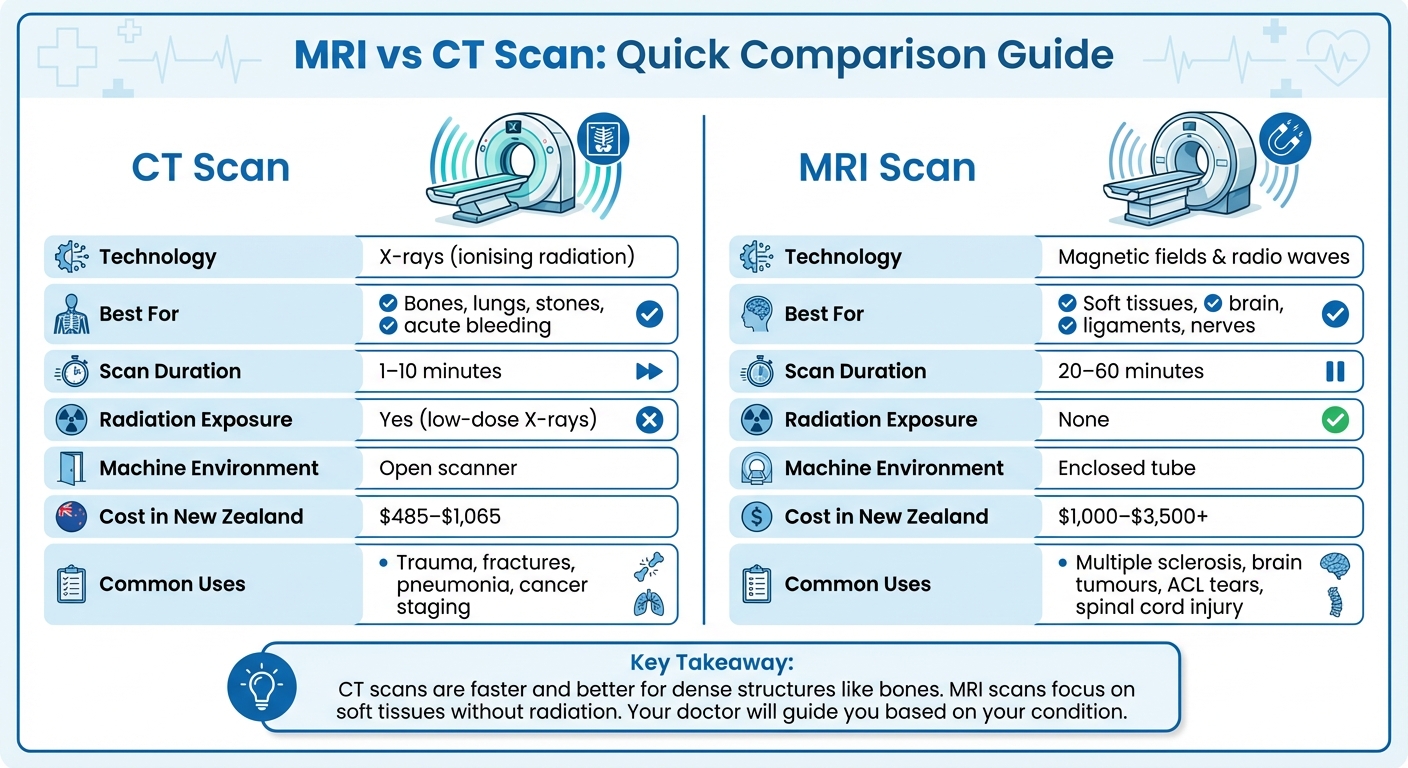
Key takeaway: CT scans are quicker and better for dense structures, while MRI scans focus on soft tissues without radiation. Your doctor will guide you based on your condition.
MRI vs CT Scan Comparison Chart: Technology, Uses, Duration, Cost and Radiation
MRI vs CT head-to-head (magnet vs X-rays)
sbb-itb-16a0ccc
How MRI Works
An MRI scanner creates images using a powerful magnetic field and low-energy radio waves. Here’s how it works: the magnetic field aligns hydrogen atoms in your body, then radio waves prompt these atoms to emit signals. A computer processes these signals to produce detailed, three-dimensional images.
Because it doesn’t use ionising radiation, MRI is safe for repeat scans, making it especially useful for monitoring conditions over time. Its ability to distinguish between soft tissues allows doctors to spot subtle differences in organs, muscles, and nerves.
Scans usually take anywhere from 15 to 90 minutes, depending on the area being examined. During the procedure, you’ll likely hear rhythmic tapping sounds, but clinics often provide earplugs or headphones for comfort. This precision in imaging is what makes MRI so effective for examining soft tissues.
What MRI Does Best
MRI excels at capturing high-resolution images of the brain, spinal cord, muscles, ligaments, tendons, and cartilage. It can even reveal nerves and pinpoint inflammation or injuries.
"MRI offers even greater detail when a more focused view is needed, especially for certain organs and soft tissues." – Memorial Sloan Kettering Cancer Center
For internal organs, MRI provides clear visuals of the heart, liver, prostate, breasts, and kidneys. There’s also a specialised technique called Magnetic Resonance Angiography (MRA), which maps blood flow through arteries and veins. MRA is often used to detect aneurysms or blockages without requiring invasive procedures.
When Doctors Order an MRI
Doctors often rely on MRI to diagnose conditions involving soft tissues or the nervous system. It’s commonly used to identify herniated discs, ligament injuries like ACL or meniscus tears, multiple sclerosis, brain tumours, strokes, and epilepsy. For sports injuries, MRI is the go-to method for assessing damage to cartilage, tendons, and joints in areas like the knees, shoulders, and hips.
"MRI is also the preferred method for evaluating cancer that may have spread to the brain or bone because it can detect subtle changes." – Dr. Richard Do, Director of MRI, Memorial Sloan Kettering Cancer Center
However, because MRI relies on a strong magnetic field, it’s crucial to inform the radiology team if you have any metallic implants, such as pacemakers, cochlear implants, or aneurysm clips, as these can pose risks during the scan. You’ll also need to remove jewellery, watches, and other metal items before the procedure.
How CT Scans Work
A CT (computed tomography) scanner blends X-ray technology with advanced computer systems to create detailed cross-sectional images of the body. The scanner itself is a ring-shaped machine equipped with an X-ray tube and detectors that rotate around you while you lie on a motorised table.
"A CT scan uses x‑rays to take detailed pictures in very fine slices through the part of your body being investigated." – Health New Zealand
As the table moves through the scanner, X-ray detectors capture images from multiple angles. These are then processed into 3D representations of bones, organs, and tissues. The process is swift, with most scans taking only seconds or minutes – much faster than an MRI. You might notice a gentle whirring sound as the machine operates.
Before the scan, you may need to fast for 2 to 4 hours, especially if a contrast dye will be used. Jewellery, dentures, glasses, and piercings should also be removed. During the scan, staying still and holding your breath briefly helps produce sharper images. If a contrast dye is involved, drinking extra water afterwards helps flush the iodine-based solution from your system. This combination of speed and precision makes CT scans a powerful diagnostic tool.
What CT Scans Do Best
CT technology shines when it comes to imaging dense structures and acute conditions. It is particularly effective for examining bones, making it the go-to option for identifying complex fractures and joint issues. Additionally, CT scans excel at detecting problems in lung tissue and spotting kidney or gallstones.
"CTs are good at looking at ‘bones and stones’ – like kidney stones and gallstones." – Dr. Albert Parlade, Radiologist, Cleveland Clinic
Often described as a "3D X-ray", CT scans provide much more detail and depth than standard X-rays. This makes them invaluable for surgeons, especially when planning procedures that require precise 3D reconstructions of bones to address complex injuries. Beyond bones, CT scans deliver clear images of internal organs like the liver, pancreas, and kidneys, helping doctors make accurate and timely diagnoses.
When Doctors Order a CT Scan
The strengths of CT scans make them essential in specific medical situations. In emergencies, such as after a car accident, CT scans are often ordered to quickly diagnose life-threatening issues like internal bleeding, organ damage, or brain injuries. Their speed is a critical advantage compared to MRI scans, which take longer.
"CT scans are fast and often used when doctors need to make quick decisions, such as in emergency situations." – Dr. Oguz Akin, Service Chief in Body Imaging, MSKCC
CT scans are also frequently used to diagnose acute conditions like appendicitis, strokes, pneumonia, and internal bleeding. They play a key role in cancer care, helping to stage the disease and monitor the effectiveness of treatments. In New Zealand, the average environmental background radiation dose is about 2 mSv per year, while a typical head CT scan exposes you to approximately 1–3 mSv. While CT scans involve more radiation than standard X-rays, their diagnostic benefits generally outweigh the risks in most cases.
MRI vs CT Scan: Main Differences
MRI and CT scans rely on different technologies, each excelling in specific areas, which influences what they’re best suited for.
Side-by-Side Comparison
The primary distinction lies in how these scans work. CT scans use X-rays to create cross-sectional images of the body, while MRI uses magnetic fields and radio waves to align water molecules in tissues. This difference shapes their strengths.
CT scans are excellent for capturing detailed images of dense structures like bones, making them a go-to for identifying fractures or lung issues. On the other hand, MRI is better at distinguishing between soft tissues, making it ideal for diagnosing conditions like ligament injuries, brain tumours, and spinal cord problems.
Here’s a quick comparison of their features:
| Feature | CT Scan | MRI Scan |
|---|---|---|
| Technology | X-rays (ionising radiation) | Magnetic fields and radio waves |
| Best For | Bones, lungs, stones, acute bleeding | Soft tissue, brain, ligaments, nerves |
| Scan Duration | 1–10 minutes | 20–60 minutes |
| Radiation Exposure | Low-dose X-rays | None |
| Machine Environment | Open scanner | Enclosed tube |
| Common Uses | Trauma, fractures, pneumonia, cancer staging | MS, brain tumours, ACL tears, spinal cord injury |
"MRI offers even greater detail when a more focused view is needed, especially for certain organs and soft tissues." – Dr. Richard Do, Director of MRI, Memorial Sloan Kettering Cancer Center
Radiation and Safety
Safety is another critical factor when choosing between MRI and CT scans.
MRI is radiation-free, which makes it a safer choice for children, pregnant women, and individuals requiring multiple scans over time. CT scans, while involving low-dose X-rays, are generally considered safe, with their diagnostic benefits outweighing potential risks in most cases.
However, MRI comes with its own precautions. The strong magnetic field can interfere with or move certain metal implants. Patients with pacemakers, cochlear implants, or older aneurysm clips must be carefully evaluated before undergoing an MRI. Many modern implants are MRI-compatible, but it’s vital to inform the radiology team of any metal in your body.
Pregnancy adds another layer of consideration. For pregnant patients, MRI or ultrasound is often preferred to avoid exposing the baby to radiation. CT scans are only used during pregnancy when absolutely necessary, and the benefits clearly outweigh the risks to the fetus.
For those who experience claustrophobia, the enclosed design of an MRI machine can be intimidating. If this is a concern, options like "wide-bore" MRI machines or mild sedatives may help make the process more comfortable. These factors are important for patients in New Zealand as they weigh the pros and cons of each imaging method.
Costs for MRI and CT Scans in New Zealand
Knowing the costs of medical imaging is crucial, whether you’re paying privately or relying on ACC support.
MRI scans tend to be more expensive than CT scans because of the advanced technology and the time required for scanning. If you’re considering a private MRI, expect to pay between $1,000–$1,400 for simpler scans like knees or shoulders. For more complex areas, such as the brain, spine, or abdomen, costs can range from $1,300–$3,500+. If a contrast agent (a dye used to improve image quality) is needed, this can add $1,000 or more to your bill.
CT scans are generally easier on the wallet. A head CT scan might cost you around $600, while scans of the chest, abdomen, or pelvis are closer to $1,065. A sinus CT scan is one of the more affordable options, with prices starting at $485.
ACC funding can drastically reduce your costs. If your scan is required due to an accident, ACC often covers the entire cost at accredited clinics, leaving you with no out-of-pocket expenses. In 2023, ACC allocated nearly $130 million to medical imaging, with MRI scans alone accounting for around $100 million. However, if you’re paying privately without ACC support, you might end up paying 17% to 53% more than the rates ACC reimburses clinics.
For those with a Community Services Card or Gold Card, it’s worth asking your radiology provider about discounts – some clinics offer a 10% reduction. Always request a written quote before your appointment, but keep in mind that the final cost might increase if the radiologist decides additional views or contrast are necessary.
These details can help you make informed decisions when looking for cost-effective radiology services in New Zealand.
Finding Radiology Services in New Zealand
If you’ve got a referral from your GP or specialist – which is required for both MRI and CT scans in New Zealand – the next step is choosing the right clinic. Radiology Clinics NZ makes this easier by providing a searchable directory of imaging providers throughout the country.
With your referral ready, you can use the directory to search by location, helping you find clinics nearby. Each clinic profile includes key details like addresses, contact information, the types of services offered (MRI, CT, ultrasound, etc.), and even user reviews to guide your decision.
For both public and private imaging needs, you’ve got options. Alongside Radiology Clinics NZ, Health New Zealand (Te Whatu Ora) also offers a directory that covers 16 regions, including Auckland, Canterbury, Waikato, Wellington, and Otago. On the private side, providers such as Auckland Radiology Group and Bay Radiology deliver specialised imaging services in specific regions. To improve access, some clinics even offer mobile PET-CT scanners that travel to different parts of the country.
When deciding on a provider, consider factors like wait times and how close the clinic is to you. It’s also important to confirm that the clinic offers the specific scan you need, as some focus on particular types of imaging or body areas. Many directories, like the one from Radiology Clinics NZ, include map features to help you plan your trip.
Before booking your appointment, let the clinic know if you have any metallic implants or experience claustrophobia. Many facilities now have wide-bore MRI machines with better lighting to help patients feel more at ease, and your GP can prescribe a sedative if necessary.
Conclusion
MRI and CT scans serve different diagnostic purposes, and knowing their main differences can help you and your doctor decide which test suits your situation best. CT scans are quick and excel at capturing details of bones, making them ideal for emergencies like detecting fractures, kidney stones, or lung conditions. On the other hand, MRI scans are better for examining soft tissues like the brain, spinal cord, ligaments, and tendons. While they take longer and require a more patient process, they don’t use ionising radiation. These distinct strengths help guide the decision-making process.
The type of scan you need depends on your specific condition. For soft tissue issues or when repeated imaging is necessary – particularly for children or during pregnancy – MRI is often the preferred choice because it avoids radiation exposure.
In New Zealand, you can access either type of scan through public or private healthcare providers with a referral. It’s important to share relevant details with your doctor, such as whether you have metal implants (for MRI) or a possible pregnancy (for CT scans). If claustrophobia is a concern, talk to your doctor about sedation options. These discussions ensure the imaging process is both safe and effective.
Ultimately, working closely with your healthcare provider is key to selecting the right test. Both MRI and CT scans are powerful diagnostic tools, offering the detailed images needed for accurate diagnoses and effective treatment plans.
FAQs
Do I need a referral for an MRI or CT scan in New Zealand?
Yes, in New Zealand, you’ll generally need a referral from a qualified healthcare professional – like a GP, specialist, or medical practitioner – for an MRI or CT scan. It’s important to discuss your situation with your doctor to ensure the imaging test recommended is the best option for your specific needs.
Can I have an MRI if I have metal implants or a pacemaker?
Having metal implants or a pacemaker can sometimes impact your ability to have an MRI. While many implants are designed to be safe for MRI scans, some, such as specific pacemakers, might carry risks. It’s crucial to let your healthcare provider and the MRI technologist know about any implants you have. They can check whether your implant is compatible with MRI or suggest other imaging options if necessary.
Will ACC cover the cost of my MRI or CT scan?
Yes, ACC might cover the cost of an MRI or CT scan in New Zealand, but it depends on specific conditions. For MRIs, your GP needs to be accredited under the GP Referred MRI (GPMRI) programme. CT scans are generally covered for accidental injuries, particularly when done in public hospitals, where they are usually free. However, private providers may charge higher fees, and whether ACC covers these costs depends on the reason for the scan and the referral process.
Related Blog Posts
- 10 Questions to Ask Before Your First Radiology Scan
- Understanding Radiology Wait Times in NZ: A Guide
- Top Clinics Offering High-Resolution Imaging in NZ
- Top PET Scan Clinics in NZ

Author: Dr. Mark Bekhit
Radiologist and reviewer for Radiology Clinics NZ medical-adjacent directory and guide content.
Editorial and medical context
This guide is published by Radiology Clinics NZ and reviewed for medical-adjacent accuracy by Dr. Mark Bekhit, radiologist. It is general information only and does not replace advice from your own doctor, referrer, clinic, or insurer.
Last editorial review: 2026-06-16.
Leave a Reply